Tooth Extraction Healing Stages: Day by Day Visual Guide
Written & Researched By
Editorial Team · product research desk
Sources: ADA guidelines, Mayo Clinic, PubMed, peer-reviewed dental literature
Published: May 26, 2026
Short answer: Tooth extraction healing follows a predictable pattern — from blood clot formation (day 1) to granulation tissue (days 3-7) to tissue closure (weeks 2-3) to complete bone remodeling (months 3-6). Knowing what each stage looks like helps you distinguish normal healing from complications that need attention.
One of the biggest sources of anxiety after extraction is looking at the hole in your gums and wondering, “Is this normal?” The socket goes through dramatic color and texture changes that can look alarming if you don’t know what to expect. This guide walks through each healing stage so you know exactly what you’re seeing.
This page is the visual-stage companion. The calendar-style writeup is Tooth Extraction Recovery Day by Day. Wisdom-tooth staging is Wisdom Teeth Recovery Timeline Day by Day. The non-medical comfort checklist is Day-by-Day Comfort Routine Checklist.
Stage 1: Blood Clot Formation (Day 0-2)
What’s Happening
Within minutes of your tooth being removed, blood fills the empty socket and begins clotting. This blood clot is the foundation of your entire healing process — it protects the exposed bone and nerve endings, provides a scaffold for new tissue growth, and prevents bacteria from reaching the bone.
What You’ll See
- Immediately after: The socket fills with dark red blood. Your dentist places gauze for you to bite on, which applies pressure to encourage clot formation.
- Hours 2-6: The blood darkens and begins solidifying into a jelly-like clot. The area looks deep red to dark burgundy.
- Day 1: A stable, dark red clot fills the socket. The surrounding gum tissue is swollen and may appear pink to dark pink. Some blood-tinged saliva is normal.
- Day 2: The clot is darker, often appearing dark red to almost black. Surrounding tissue may be more swollen than day 1 (swelling typically peaks at 48-72 hours).
What’s Normal vs. Concerning
| Normal | Concerning |
|---|---|
| Dark red to blackish clot in socket | Empty socket — can see white bone |
| Mild oozing, blood-tinged saliva | Heavy, active bleeding after 6+ hours |
| Increasing swelling through day 2 | Severe swelling that impairs breathing |
| Dull, manageable pain | Intensifying, radiating pain |
For detailed information on the blood clot stages, read our complete blood clot stages guide.
Stage 2: Inflammatory Phase (Days 2-4)
What’s Happening
Your immune system is working at full capacity. White blood cells flood the area to fight bacteria and clear debris. New tiny blood vessels begin growing into the clot (a process called angiogenesis). This stage is essential — inflammation is your body’s healing mechanism, not a problem to eliminate.
What You’ll See
- Swelling peaks around 48-72 hours after extraction. Your cheek or jaw may be noticeably puffy.
- The clot surface may develop a yellowish or grayish film. This is fibrin — a protein involved in clot stabilization — and it’s completely normal.
- Surrounding gums appear red, puffy, and possibly bruised-looking (especially after surgical extractions).
- Slight color changes at the clot surface, moving from pure dark red toward a more mottled appearance.
Pain Expectations
This is typically when pain is most noticeable. Days 2-3 are often described as the worst for discomfort, which then gradually improves. If pain is getting worse instead of better by day 4, this is the critical warning sign for dry socket.
Stage 3: Granulation Tissue Formation (Days 4-7)
What’s Happening
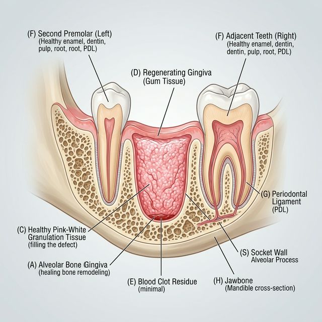
This is one of the most important healing stages. Granulation tissue — a soft, moist tissue rich in new blood vessels and collagen — begins replacing the blood clot from the edges of the socket inward. This tissue is your body’s wound-healing material, creating the foundation for new gum tissue and eventually new bone.
What You’ll See
- A white or creamy-white film begins covering the dark clot surface. This is the granulation tissue growing over the clot, and it often alarms patients who mistake it for infection or pus.
- The socket appears lighter, transitioning from dark red/black to pinkish-white.
- The edges of the gum may start pulling inward slightly as new tissue contracts.
- Swelling decreases noticeably from day 4 onward.
The Key Distinction
White tissue forming over the clot surface = normal granulation tissue (healthy healing).
White bone visible at the bottom of an empty-looking socket + severe pain = dry socket (needs treatment).
The presence or absence of pain is the best indicator. Granulation tissue formation is not painful — if anything, you should feel improvement. For a deeper dive into recognizing healthy white tissue, see our granulation tissue guide.
Stage 4: Epithelial Closure (Weeks 1-3)
What’s Happening
Epithelial cells — the type that form your gum surface — begin migrating across the granulation tissue from the socket edges toward the center. Think of it like a pond freezing over: the edges close first, gradually working inward until the surface is sealed.
What You’ll See
- The socket opening shrinks visibly, getting smaller each day.
- The tissue color shifts from white/pink granulation tissue to a more normal pink that increasingly matches your surrounding gum tissue.
- The hole becomes shallower as the bottom fills in with tissue from below while the surface closes from the sides.
- By week 2-3, the surface may appear fully or nearly closed for a simple extraction. Wisdom tooth sites take longer due to their larger size.
Pain Expectations
Pain should be minimal to nonexistent by this stage. Occasional sensitivity when eating or brushing near the site is normal, but active pain is not expected. If you’re still experiencing notable pain at 2 weeks, contact your dentist to rule out infection. See our guide on signs of infection after extraction for what to watch for.
Stage 5: Tissue Maturation (Weeks 3-8)
What’s Happening
The gum surface has closed, and the tissue underneath is maturing and strengthening. Collagen fibers reorganize for durability. The indentation where the tooth was gradually fills in, though it may never completely level with the surrounding bone.
What You’ll See
- The surface looks like normal gum tissue — pink, smooth, and healthy.
- A slight depression may be visible or palpable where the tooth was, especially for larger teeth like molars.
- No pain, swelling, or sensitivity should be present.
Stage 6: Bone Remodeling (Months 2-6)
What’s Happening
Beneath the healed gum surface, your jawbone is undergoing remodeling. New bone fills in the socket from the bottom up, eventually replacing the extraction site with solid bone. This process is slower and invisible from the outside.
What You’ll Notice
- The indentation or gap gradually fills in with firm tissue as bone density increases.
- By 4-6 months, the bone has remodeled enough for procedures like dental implants if planned.
- Some bone loss at the surface is normal after extraction, which is why dentists sometimes place a bone graft at the time of extraction if an implant is planned later.
Healing Timeline at a Glance
| Stage | Timeframe | What You See | Pain Level |
|---|---|---|---|
| Blood clot | Days 0-2 | Dark red/black filling in socket | Moderate, managed with medication |
| Inflammation | Days 2-4 | Swelling peaks, yellowish film on clot | Worst days, then improving |
| Granulation tissue | Days 4-7 | White/pink tissue covering clot | Noticeably improving |
| Surface closure | Weeks 1-3 | Socket opening shrinks, tissue pinks | Minimal to none |
| Tissue maturation | Weeks 3-8 | Looks like normal gum | None |
| Bone remodeling | Months 2-6 | Invisible (beneath surface) | None |
Factors That Slow Healing
Several factors can extend these timelines:
- Smoking — the single largest controllable factor in delayed healing. Nicotine restricts blood flow to the site.
- Poor nutrition — your body needs protein, vitamin C, and zinc to build new tissue. Under-eating during recovery slows the process.
- Dry socket — if the blood clot dislodges in stage 1-2, healing essentially restarts after treatment.
- Infection — bacterial infection interrupts the healing sequence and requires antibiotics.
- Complex extractions — surgical removal of impacted teeth creates a larger wound that takes longer to heal at each stage.
- Medical conditions — diabetes, autoimmune disorders, and medications like steroids or blood thinners can delay healing.
When to Call Your Dentist
During any healing stage, contact your dental provider if you notice:
- Pain that worsens instead of gradually improving after day 3
- Heavy bleeding that restarts after initially stopping
- Fever above 101°F (38.3°C)
- Pus or foul-smelling discharge from the socket
- Numbness that persists beyond 8 hours after the procedure
- Difficulty opening your mouth that gets worse instead of better after day 3
- A socket that looks empty with visible bone instead of being filled with tissue
How to Support Each Healing Stage
Stage 1-2 (Clot protection): Rest, bite on gauze as directed, avoid straws and smoking, sleep with your head elevated, apply ice packs 20 minutes on/20 off.
Stage 3-4 (Tissue growth): Start gentle warm salt water rinses (1/2 teaspoon salt in 8 ounces warm water), eat soft foods rich in protein, avoid poking the site with your tongue.
Stage 5-6 (Maturation): Resume normal brushing (gently around the site), gradually return to your regular diet, and attend your follow-up appointment.
For a day-by-day recovery roadmap with specific instructions, see our tooth extraction recovery guide.
Frequently Asked Questions
What does an infected extraction site look like?
An infected site typically shows redness and swelling that gets worse after day 3-4 (instead of improving), possible pus or cloudy discharge, and the surrounding gum tissue may appear darker red or purplish. Infection is often accompanied by fever, worsening pain, and a foul taste. Healthy healing tissue is pale pink to white; infected tissue looks angry and inflamed.
Is it normal for the extraction site to be black?
A very dark red or blackish appearance in the first 2-3 days is usually just a dense blood clot — this is normal and healthy. The color darkens as the blood in the clot deoxygenates. If the dark color is accompanied by foul odor, pus, or increasing pain, it could indicate a problem.
How long does the hole take to close after extraction?
Surface closure typically takes 2-3 weeks for simple extractions and 4-6 weeks for surgical wisdom tooth removals. The underlying bone takes 3-6 months to fully remodel. You may feel a shallow indentation at the site for several months, which is normal. Socket closing is not the same as neighboring teeth moving; that later distinction is explained in teeth shifting after tooth extraction.
Why is my extraction site still open after 2 weeks?
For larger teeth or surgical extractions, 2 weeks may not be enough time for complete surface closure. This is especially true for wisdom teeth. If the open area is getting smaller, there’s no pain, and there’s no signs of infection, healing is likely progressing normally — just slowly. If in doubt, see your dentist for a quick check.
Can I speed up extraction healing?
You can’t dramatically accelerate biology, but you can avoid slowing it down. Don’t smoke, eat protein-rich foods, stay hydrated, follow your dentist’s rinse instructions, and get adequate sleep. These won’t make healing faster than normal — but they prevent the delays that many patients experience from not following these basics.
Key Takeaway
Your extraction socket goes through dramatic-looking changes during healing, and nearly all of them are normal. The most reliable indicator of healthy healing isn’t what the socket looks like — it’s your pain trend. If pain is improving daily from day 3 onward, you’re almost certainly healing well. If pain reverses course and gets worse, that’s the signal to call your dentist. Trust the process, follow your post-op instructions, and let your body do what it’s designed to do.
Written and researched by the editorial team. Sources: ADA guidelines, Mayo Clinic, peer-reviewed dental literature.
Sources: Journal of Oral and Maxillofacial Surgery; American Association of Oral and Maxillofacial Surgeons; American Dental Association.