Tooth Extraction Blood Clot Stages: What Your Socket Should Look Like
Written & Researched By
Editorial Team · product research desk
Sources: ADA guidelines, Mayo Clinic, PubMed, peer-reviewed dental literature
Published: May 25, 2026
Short answer: The blood clot in your extraction socket changes appearance as it heals. It starts as a dark red jelly-like mass on day 0, transitions to a lighter pinkish-white (granulation tissue) around days 3-5, gets covered by gum tissue between days 7-14, and the socket closes completely within 3-4 weeks. Each stage looks different, and knowing what to expect prevents unnecessary worry.
One of the most common concerns after a tooth extraction is looking at the socket and not knowing whether what you see is normal. The color changes, the texture changes, and sometimes what’s happening in there looks alarming when it’s actually healthy healing. Here’s a visual guide to each stage.
Stage 1: Blood Clot Formation (Day 0, First Few Hours)
What’s happening
Immediately after the tooth is removed, blood fills the empty socket. Within 15-30 minutes of sustained pressure (biting on gauze), platelets in the blood begin aggregating and forming a clot. This clot fills the socket like a biological plug.
What it looks like
- Color: Dark red, like a blob of dark jelly
- Texture: Soft and slightly raised, sitting in or slightly above the socket opening
- Size: Fills the entire socket opening
- You may see blood-tinged saliva for the first 4-6 hours — this is normal oozing, not active bleeding
What it’s doing
The clot serves as a protective cover over the exposed bone and nerve endings at the bottom of the socket. Without this clot, those structures are exposed to air, food, and bacteria, which causes the intense pain of dry socket.
The clot also provides a framework. New tissue will grow through and on top of this clot structure. Think of it as the scaffolding that the rest of the healing process builds on.
How to protect it
This stage is the most vulnerable. The clot is fresh and can be dislodged easily.
- Bite on gauze for 30-45 minutes with firm, constant pressure
- Do not rinse, spit, or use straws for 24 hours
- Do not smoke — the suction and chemicals both threaten clot stability
- Eat only cool, soft foods on the opposite side of your mouth
- Sleep with your head elevated on a wedge pillow to reduce blood pressure at the site
Stage 2: Clot Maturation (Days 1-2)
What’s happening
The fresh clot stabilizes and begins organizing. White blood cells infiltrate the clot to clean up debris and fight bacteria. Your body’s inflammatory response peaks during this window, which is why swelling and pain are at their worst.
What it looks like
- Color: Still dark red, but may start developing lighter edges. Some patients notice a whitish or yellowish film on the surface — this is fibrin, a protein involved in the clotting process, and it’s completely normal
- Texture: Firmer than day 0, slightly less jelly-like
- The surrounding gum: Red, swollen, tender to touch
Common concern: “Is that white stuff on my clot normal?”
Almost always yes. The white or yellowish material you see on the surface of the clot is fibrin. It’s part of the clot structure and a normal part of wound healing. It is not pus. Pus is thicker, often greenish, and accompanies other symptoms like fever and increasing pain. Fibrin is thin, filmy, and sits on the clot surface.
Stage 3: Granulation Tissue Formation (Days 3-5)
What’s happening
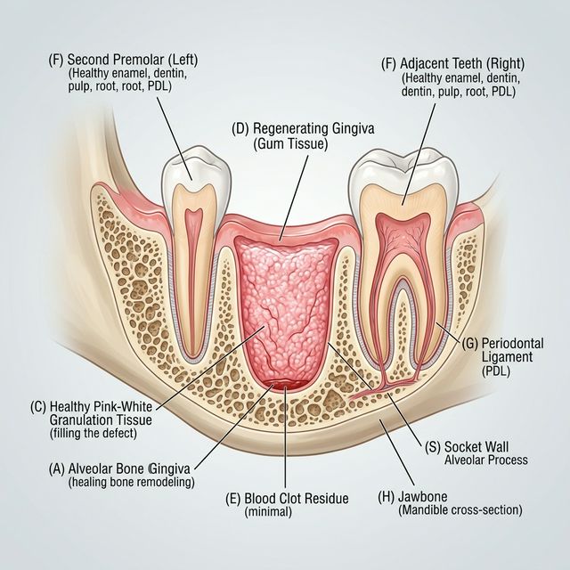
This is the most dramatic visual change. The blood clot is being gradually replaced by granulation tissue — a new type of tissue that your body builds specifically for wound repair. New blood vessels sprout into the area (a process called angiogenesis), bringing nutrients and oxygen needed for healing.
What it looks like
- Color: Shifting from dark red to pinkish, pinkish-white, or creamy white. The socket may look lighter than you expect.
- Texture: Soft, slightly bumpy or pebbly. Some patients describe it as looking like a wet, pink marshmallow. It’s fragile — don’t poke or prod it.
- Size: The socket is starting to shrink as the tissue fills in from the edges and bottom
For a detailed look at granulation tissue and what it means, read granulation tissue after tooth extraction: what it is and why it matters.
Common concern: “My socket is turning white. Is this dry socket?”
Probably not. Dry socket has specific accompanying symptoms: severe, worsening pain that radiates to the ear or temple, a bad taste, and OTC pain meds that don’t help. If the socket is turning whitish-pink but your pain is improving daily, what you’re seeing is healthy granulation tissue. For a full comparison, see dry socket symptoms vs normal healing.
Stage 4: Tissue Coverage (Days 5-10)
What’s happening
The gum tissue at the edges of the socket is migrating inward, gradually covering the granulation tissue beneath. The surface of the socket transitions from an open wound to a closed surface. Deeper in the socket, early bone formation is beginning.
What it looks like
- Color: Pinkish-red gum tissue growing in from the edges, with lighter tissue visible in the center
- Texture: Smoother than the granulation tissue stage. The bumpy, pebbly appearance gives way to more uniform tissue
- Size: The opening is visibly smaller than it was a week ago
- A shallow depression or indentation is normal — the socket doesn’t fill all the way to the surface in this stage
What to watch for
This is typically a low-risk stage. By now, the chances of developing dry socket are very low (dry socket almost always begins between days 2-4). Continue being gentle when brushing near the area and rinsing after meals with an alcohol-free rinse like CloSYS or warm salt water.
Stage 5: Surface Closure (Days 10-21)
What’s happening
The gum tissue fully covers the extraction site. Beneath the closed surface, the socket continues filling with new bone tissue. The soft tissue on top is still maturing and strengthening.
What it looks like
- Color: Pink, matching the surrounding gum tissue. May be slightly more red or shiny than the adjacent gums for a few more weeks
- Texture: Smooth, continuous gum surface. The site may look slightly indented compared to the surrounding tissue
- No open wound is visible
What you can do
- Resume normal brushing and flossing in the area
- Eat normally — hard and crunchy foods are fine now for most patients
- The indentation will gradually fill in over the coming months as bone remodels underneath
Stage 6: Bone Remodeling (Months 1-6)
What’s happening
This is the longest stage but the least noticeable. Woven bone (hastily built structural bone) is gradually replaced by lamellar bone (mature, organized bone). The socket fills in completely from the bottom up.
What it looks like
From the outside, nothing. The gum surface looks normal. You may feel a slight depression with your tongue for the first month or two, but it gradually levels out. By 4-6 months, an X-ray would show fully remodeled bone filling the socket.
This stage matters most for patients planning dental implants, since an implant requires sufficient bone volume. Your dentist may have placed a bone graft at the time of extraction to preserve the socket shape for a future implant.
Visual Quick Reference
| Stage | Days | Color | Texture | Pain |
|---|---|---|---|---|
| Blood clot formation | Day 0 | Dark red | Jelly-like, soft | Moderate |
| Clot maturation | Days 1-2 | Dark red + white film (fibrin) | Firmer, stable | Peak |
| Granulation tissue | Days 3-5 | Pinkish-white, creamy | Bumpy, pebbly, soft | Improving |
| Tissue coverage | Days 5-10 | Pink edges closing inward | Smoother, shrinking opening | Mild |
| Surface closure | Days 10-21 | Pink, matching gums | Smooth, continuous | Minimal to none |
| Bone remodeling | Months 1-6 | Normal gum color | Normal gum texture | None |
What the Stages Look Like When Something Goes Wrong
Dry socket
The clot is missing. Instead of a dark red or pinkish-white filling, the socket looks hollow with whitish or grayish bone visible at the bottom. Accompanied by severe pain starting on day 2-4 that radiates to the ear or temple. Read the dry socket symptoms guide for the full picture.
Infection
The socket tissue looks red, angry, and swollen. You may see thick, yellowish-green pus (not to be confused with thin, whitish fibrin). Accompanied by fever, increasing pain, and possibly swollen lymph nodes. See infection signs after tooth extraction.
Bone spicule
A small, hard, white fragment poking through the gum tissue. This is a piece of bone that broke off during extraction and is working its way to the surface. Usually uncomfortable but not dangerous — your dentist can remove it in a quick visit. Read more about bone spicules after tooth extraction.
Frequently Asked Questions
What color should a healing tooth extraction look like?
Dark red on days 0-2 (blood clot), transitioning to pinkish-white on days 3-5 (granulation tissue), then gradually matching the pink of your surrounding gum tissue as the surface closes over days 7-21. The changing colors are a sign of healthy progression, not a reason for concern.
Is it normal for the extraction site to be white?
Yes, in most cases. White or pinkish-white tissue appearing on days 3-5 is granulation tissue — a healthy sign that your body is building new tissue. However, if the white appearance is accompanied by severe, worsening pain and visible bone at the bottom of the socket, it may indicate dry socket.
How do I know if my blood clot fell out?
If the clot dislodges, you may taste blood or notice the socket looks empty. The most telling sign is a sudden worsening of pain on day 2-4 that is distinctly more severe than day 1. You may see bone at the bottom of the socket instead of a dark red filling.
Can I see the blood clot in my extraction socket?
Yes, usually. Look in a mirror with a flashlight. In the first 1-3 days, you should see a dark red mass filling the hole where the tooth was. As healing progresses, the appearance changes (see the stages above), but the socket should always look filled with tissue, not hollow.
How long does the blood clot stay in the socket?
The blood clot itself is gradually replaced by granulation tissue starting around day 3. By day 7-10, the original clot has been almost entirely replaced by new tissue. You don’t lose the clot — it transforms. The risk of dry socket (from clot dislodgement) is highest in the first 3-4 days.
Key Takeaway
Your extraction socket goes through dramatic visual changes during healing — dark red, whitish film, pinkish-white, and finally normal pink gum tissue — and all of these changes are normal. The key indicator is your pain trend, not the color of the socket. If pain is improving daily from day 3 onward, what you’re seeing in the socket is almost certainly healthy healing. If pain is worsening after day 2-3, regardless of what the socket looks like, call your dentist.
Medical Disclaimer: This article is for informational purposes only and does not replace professional dental evaluation. If you are concerned about the appearance of your extraction site or experiencing unusual symptoms, contact your dentist or oral surgeon for a clinical assessment.
Written and researched by the editorial team. Sources: ADA guidelines, Journal of Oral and Maxillofacial Surgery, American Association of Oral and Maxillofacial Surgeons, Mayo Clinic.